This is used to visualize the position of a cloned gene on a eukaryotic chromosome.
The cells are treated with a fixative. They are attached to a glass slide and incubated with RNAse and NaOH to degrade RNA and denature the DNA. The chromosome exposes the segments of DNA. A sample of the cloned gen is labelled and applied to the chromosome preparation. Hybridization occurs between the labelled cloned gene and its chromosomal copy. This results in a dark spot on autoradiography if the cloned gene was radioactively labelled. The position of this spot will indicate the location of the cloned gene on the chromosome (Figure 9.26).
Figure 9.26 Fluoresence in situ hybridization
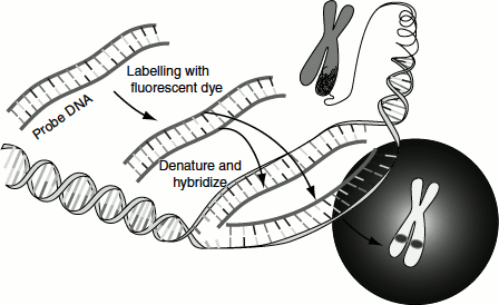
Fluorescent in situ hybridization (FISH)
A fluorescently labelled cloned gene is used as probe and is hybridized to chromosome preparation as described before. This method is also useful for characterizing chromosomal rearrangements and for analysing microdeletions.